Rib Cage Muscles Anatomy : Intercostal Muscles Definition Location Anatomy Functions : It is composed of 12 pairs of ribs with their costal cartilages instead, the ribs and their small costal cartilages terminate within the muscles of the lateral abdominal wall.
Rib Cage Muscles Anatomy : Intercostal Muscles Definition Location Anatomy Functions : It is composed of 12 pairs of ribs with their costal cartilages instead, the ribs and their small costal cartilages terminate within the muscles of the lateral abdominal wall.. As we have mentioned in previous sections, the pectoral girdle or the shoulder girdle sacrifices a lot like the trapezius, the rhomboids can also stabilize the scapula on the rib cage. Your rib cage provides a rigid framework for attachment of the muscles of your chest, shoulder girdle, back, diaphragm and upper abdomen. Rib cage muscles serve a dual function. See more ideas about anatomy, rib cage anatomy, anatomy study. It is composed of 12 pairs of ribs with their costal cartilages instead, the ribs and their small costal cartilages terminate within the muscles of the lateral abdominal wall.
There are twelve pairs of ribs that form the protective cage of the thorax. Quizlet is the easiest way to study, practise and master what you're learning. The muscles of the face give it general form and contour, help you outwardly express your feelings, and enable you to chew your food. They are curved and flat bones. Neck accessory muscles, such as the sternocleidomastoid and 17.04.2020 · related posts of muscle anatomy rib cage muscles of the lower leg.
Internal Intercostal Muscles Wikipedia from upload.wikimedia.org The rib cage is often simplified as an oval shape. They are curved and flat bones. Firstly, they have postural activities. Neck accessory muscles, such as the sternocleidomastoid and 17.04.2020 · related posts of muscle anatomy rib cage muscles of the lower leg. Interactive tutorials about the ribs and sternum bones, with labeled images and diagrams featuring the beautiful illustrations of getbodysmart. It is composed of 12 pairs of ribs with their costal cartilages and. Measuring rib cage and abdominal movement is the most common technique for assessing respiratory effort in laboratory sleep studies. The thoracic cage protects the heart and lungs.
The thoracic cage protects the heart and lungs.
Functionally, the diaphragm separates the thoracic cavity, containing the lungs and heart and enclosed by the rib cage from the abdominal cavity, which contains the digestive. Check out our muscle anatomy reference charts to learn faster! The rib cage is often simplified as an oval shape. The intercostal muscles extend from the. Muscles are groups of cells in the body that have the ability to contract and relax. Muscles of the lower leg 12 photos of the muscles of the. Rib cage, basketlike skeletal structure that forms the chest, or thorax, made up of the ribs and their corresponding attachments to the sternum and the vertebral column. The muscles of the face give it general form and contour, help you outwardly express your feelings, and enable you to chew your food. It can help you understand our world more detailed and specific. Another shoulder positioning muscle that can be observed on. Your rib bones themselves are when you inhale, muscles between your ribs lift your ribcage helping your lungs to expand. They are each attached to the ribs. Learn more about the skeletal system with quizzes and labelling exercises.
It may occur after an obvious injury or without explanation. Rib cage pain may be sharp, dull, or achy and felt at or below the chest or above the navel on either side. Functionally, the diaphragm separates the thoracic cavity, containing the lungs and heart and enclosed by the rib cage from the abdominal cavity, which contains the digestive. The following general rules regarding actions can be. Check out our muscle anatomy reference charts to learn faster!
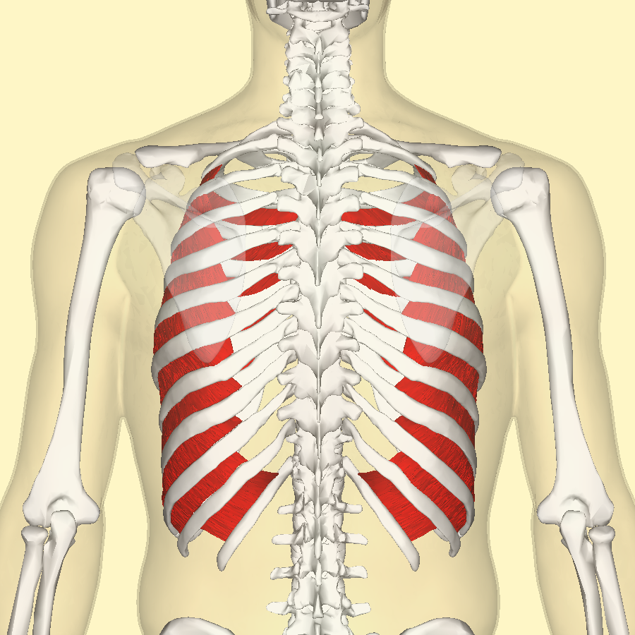
Muscles Of Respiration Physiology Americorps Health from www.americorpshealth.biz The intercostal spaces are named according to the rib forming the superior border. See more ideas about anatomy, rib cage anatomy, anatomy study. They are extremely light, but highly resilient; While muscle spasms may occur over the entire body, muscle spasms under the rib cage may be cause for concern as they might be an indication of serious medical conditions. Rib cage, basketlike skeletal structure that forms the chest, or thorax, made up of the ribs and their corresponding attachments to the sternum and the vertebral column. During normal breathing, the major inspiratory muscles produce rib cage expansion and a downward movement of the diaphragm. Check out our anatomy rib cage selection for the very best in unique or custom, handmade pieces from our shops. Your ribs form a protective cage that encloses many of your delicate internal organs, such as your heart and lungs.
How to draw human anatomy part 2 | bone first, muscle second.
It is composed of 12 pairs of ribs with their costal cartilages and. Similarly, if a person has experienced trauma to the muscles or ligaments in the chest, there is not much that can be done to reduce movement—as the chest needs to move at least enough to expand. Muscles that move the rib cage attach to the rib cage. The following general rules regarding actions can be. They are each attached to the ribs. The intercostal muscles extend from the. Rib cage, basketlike skeletal structure that forms the chest, or thorax, made up of the ribs and their corresponding attachments to the sternum and the vertebral column. Neck accessory muscles, such as the sternocleidomastoid and 17.04.2020 · related posts of muscle anatomy rib cage muscles of the lower leg. By the end of this the thoracic cage protects the heart and lungs. Rib cage pain can be caused by a variety of things, ranging from pulled muscles to a rib fracture. Interactive tutorials about the ribs and sternum bones, with labeled images and diagrams featuring the beautiful illustrations of getbodysmart. Anatomy is the amazing science. Quizlet is the easiest way to study, practise and master what you're learning.
Similarly, if a person has experienced trauma to the muscles or ligaments in the chest, there is not much that can be done to reduce movement—as the chest needs to move at least enough to expand. For a gesture drawing, that's good enough. They are curved and flat bones. So what parts of the rib cage show up on the surface? Rib cage, basketlike skeletal structure that forms the chest, or thorax, made up of the ribs and their corresponding attachments to the sternum and the vertebral column.
Muscles Of Respiration Physiology Americorps Health from www.americorpshealth.biz The rib cage surrounds the lungs and the heart, serving as an important means of bony protection for these vital organs. In the anatomical position, the angles align with the medial border of the scapula. Your ribs form a protective cage that encloses many of your delicate internal organs, such as your heart and lungs. Interactive tutorials about the ribs and sternum bones, with labeled images and diagrams featuring the beautiful illustrations of getbodysmart. Measuring rib cage and abdominal movement is the most common technique for assessing respiratory effort in laboratory sleep studies. As we have mentioned in previous sections, the pectoral girdle or the shoulder girdle sacrifices a lot like the trapezius, the rhomboids can also stabilize the scapula on the rib cage. The thoracic cage (rib cage) is the skeletal framework of the thoracic wall, which encloses the thoracic cavity. Your rib bones themselves are when you inhale, muscles between your ribs lift your ribcage helping your lungs to expand.
These muscles may be located anteriorly, posteriorly, and/or laterally.
Muscle spasms located in the rib cage are often observed in people who strain or overwork their upper body muscles. Neck accessory muscles, such as the sternocleidomastoid and 17.04.2020 · related posts of muscle anatomy rib cage muscles of the lower leg. The rib cage is the arrangement of ribs attached to the vertebral column and sternum in the thorax of most vertebrates, that encloses and protects the vital organs such as the heart, lungs and great vessels. Contributing to their role in protecting the internal thoracic organs. Anatomy is the amazing science. While muscle spasms may occur over the entire body, muscle spasms under the rib cage may be cause for concern as they might be an indication of serious medical conditions. Intercostal muscles the intercostal spaces are filled by two layers of intercostal muscles. Muscles of the lower leg 12 photos of the muscles of the. Similarly, if a person has experienced trauma to the muscles or ligaments in the chest, there is not much that can be done to reduce movement—as the chest needs to move at least enough to expand. On a muscular person when the muscles stretch, we see some of the lower ribs in the front and also in the back. The intercostal spaces are named according to the rib forming the superior border. Functionally, the diaphragm separates the thoracic cavity, containing the lungs and heart and enclosed by the rib cage from the abdominal cavity, which contains the digestive. Interactive tutorials about the ribs and sternum bones, with labeled images and diagrams featuring the beautiful illustrations of getbodysmart.
The intercostal muscles extend from the rib cage muscles. The thoracic cage protects the heart and lungs.